Life in the Fast Lane
- Highest Imaging Speed on the Market
- Gentle Sample Handling and Extremely Low Phototoxicity
- Multiview Imaging without Rotation
- High Sensitivity and Low Noise
- Ideal for Imaging Dynamic Process in Living Specimen

The MuVi-SPIM utilizes a sheet of laser light to illuminate only a thin slice of a fluorescently labeled sample. A wide-field fluorescence microscope, placed perpendicular to the light-sheet, serves to collect the fluorescence signal and images the observed region by means of a camera.  The MuVi-SPIM provides four simultaneous orthogonal views on large living specimens without the need for sample rotation. This avoids shadowing effects and facilitates long-term imaging at dramatically increased acquisition speed. Modular software concepts allow the flexible design of complex experimental layouts. 3D image data are used in real-time and directly streamed to a storage and data processing server.
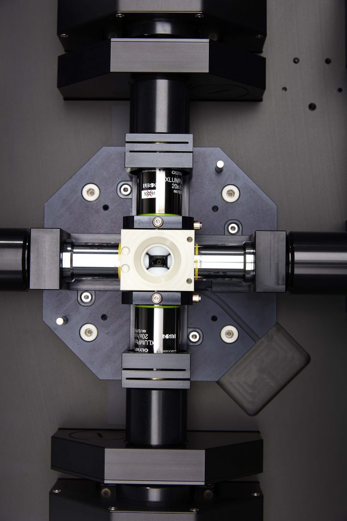
 Image of Platynereis Dumerilii.The staining is EMTB, which is a microtubule associated protein, so what you see are microtubules. Credit to: Yu-Wen Hsieh & Pavel Tomancak, MPI-CBG, Dresden, Germany
 | Laser | Laser combiner with six laser positions |
| 405, 445, 488, 515, 532, 561, 594, 642, and 685nm @ 40mW | |
| Fast modulation and high extinction | |
| Illumination optics | Two identical illumination arms |
| Chromatic correction from 440 to 660 nm | |
| Light-sheet generation by beam scanning | |
| Variable light-sheet thickness (optional) | |
| Water-dipping objective lenses (see below) | |
| Easy and intuitive alignment | |
| Detection Optics | Two identical detection arms |
| Water-dipping objective lenses (see below) | |
| Fast filter wheels with 10 positions and 50 ms between adjacent positions | |
| High-speed sCMOS cameras Hamamatsu Orca Flash 4.0 v3 with 2048 x 2048 pixels of 6.5 µm x 6.5 µm size | |
| Objective lens mounting unit | High precision mounting unit |
| Fast exchange of the objective combination unit with high precision | |
| Mounting chamber with temperature control (see below) | |
| 2 x Nikon CFI Plan Fluor 10X @0.3 NA water immersion for illumination | |
| 2 x Olympus 20X @ 1.0 NA water immersion for detection | |
| Other objective lenses on request | |
| Sample chamber and stage | Water-sealed inert PEEK plastic chamber, autoclavable and biocompatible |
| Sample supported from below for improved stability | |
| Easy access from above for sample mounting, injections, etc. | |
| Fast and precise temperature control, range 15-40°C | |
| High-precision XYZ and fast rotation stage | |
| XYZ piezo crawler stage with 100 nm resolution and 9.5 mm (X) and 1.2 mm (YZ) travel range for fast sample positioning and stack acquisition | |
| Rotation stage with > 360 degrees/s speed @ 1 degree resolution | |
| Electronics, Computer, Software | All electronic components included in the microscope housing |
| Embedded microscope software with open communication interface | |
| Open GUI control for interactive control and microscope automation | |
| Computer with 128 GB RAM, Intel Dual Six Core CPU | |
| High-speed RAID controller and 10 Gb/s Ethernet port for data streaming | |
| 8 x 4 TB local storage in RAID 0 | |
| One 40 inch monitor |
 Objectives
 Objectives Close View
 Objectives
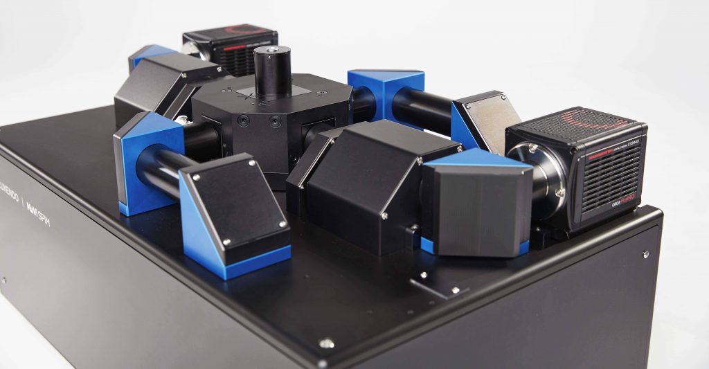
 System Overview
 Sideview
 Close View
 Close View
 System Overview